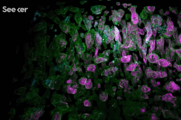
New center illuminates ‘microscopic universe within each cell’
With new advances in microscopy, Berkeley biologists are able to see the secret life of cells unfolding like never before

November 27, 2019
A new Seeker video profiles UC Berkeley's Advanced Bioimaging Center.
In a video posted online this week, Seeker, a San Francisco-based digital media network focused on science and technology, profiled UC Berkeley’s newest cutting-edge facility, the Advanced Bioimaging Center.
Run by Srigokul “Gokul” Upadhyayula, a newly arrived assistant professor-in-residence of molecular and cell biology, the center is building imaging systems that will provide real-time video of living cells for biologists who want to understand “how life works,” Upadhyayula told Seeker. He still is awestruck at the “microscopic universe inside each cell,” he said
Each of the first two imaging systems being built at the center bundle several types of imaging into a single microscope called the Multimodal Optical System with Adaptive Imaging Correction, or MOSAIC. MOSAIC includes adaptive optical lattice light-sheet microscopy, a technique developed at the Howard Hughes Medical Institute’s Janelia Research campus in the lab of Nobel Laureate Eric Betzig, who joined the Berkeley physics faculty in 2018. Betzig’s team included Upadhyayula. The technique, which they reported last year in the journal Science, allows fluorescence imaging of living cells within transparent organisms without killing them and remove the blurriness caused by surrounding cells and tissue.
Upadhyayula and the center’s co-founders — Betzig, Xavier Darzacq, Doug Koshland and Robert Tjian — hope it will become a premier visualization, data processing and analysis powerhouse with a worldwide user base and a model for other universities.
“Understanding biology and its complexity is probably one of the last human frontiers,” says Upadhyayula. “It’s the age of exploration, but instead of looking out at the galaxies, we’re looking at the galaxies inside the cells.”
More information on the Advanced Bioimaging Center, which is located in Barker Hall