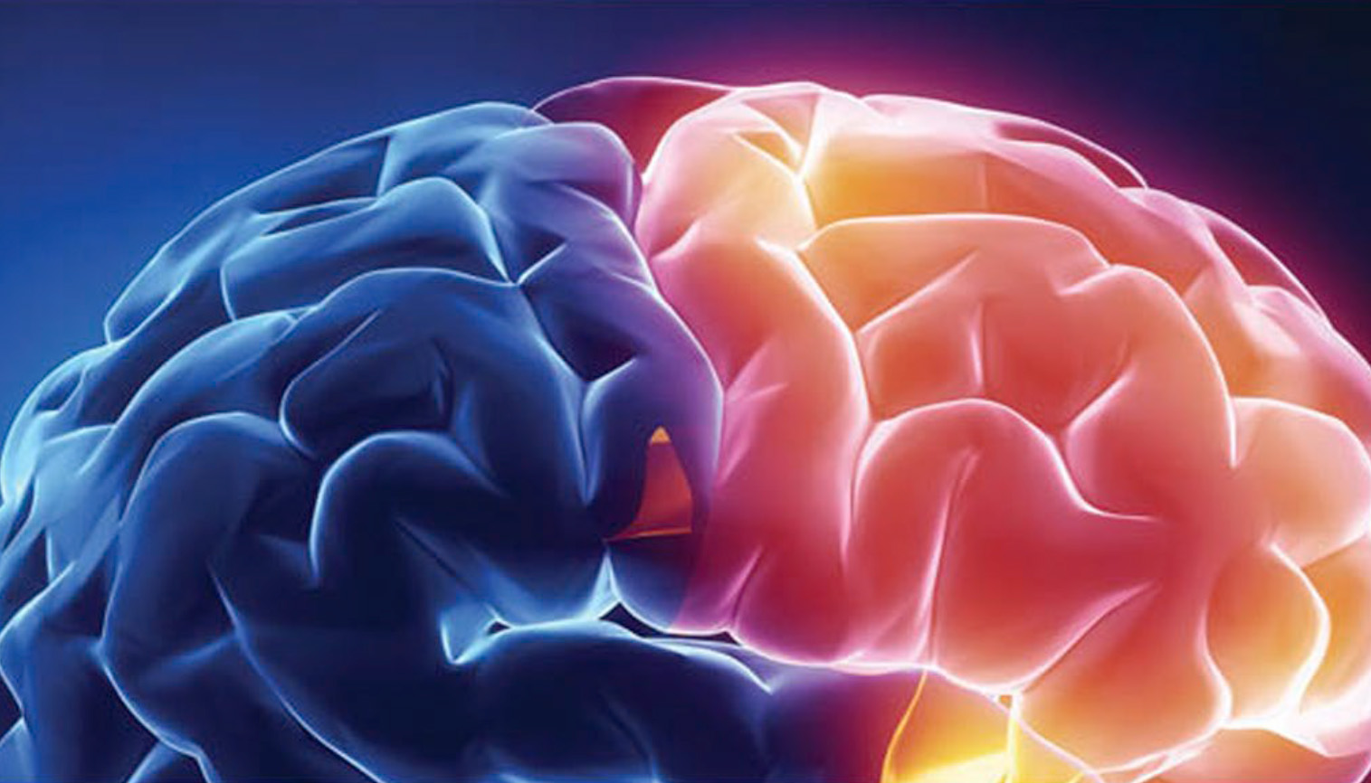
Are groovy brains more efficient?
A new study finds that the depth of small grooves in the brain's surface is linked to stronger network connectivity and better reasoning ability.
May 21, 2025
Many grooves and dimples on the surface of the brain are unique to humans, but they’re often dismissed as an uninteresting consequence of packing an unusually large brain into a too-small skull.
But neuroscientists are finding that these folds are not mere artifacts, like the puffy folds you get when forcing a sleeping bag into a stuff sack. The depths of some of the smallest of these grooves seem to be linked to increased interconnectedness in the brain and better reasoning ability.
In a study published May 19 in The Journal of Neuroscience, University of California, Berkeley, researchers show that in children and adolescents, the depths of some small grooves are correlated with increased connectivity between regions of the brain — the lateral prefrontal cortex and lateral parietal cortex — involved in reasoning and other high-level cognitive functions.
The grooves may actually bring those areas closer together in space, shortening the connections between them and speeding communications.
The implication, the researchers say, is that variability in these small grooves, which are called tertiary sulci (pronounced sul’-sigh), may help explain individual differences in cognitive performance, and could serve as diagnostic indicators or biomarkers of reasoning ability or neurodevelopmental disorders.
“The impetus for this study was having seen that sulcal depth correlated with reasoning across children and adolescents,” said Silvia Bunge, professor of psychology and a member of UC Berkeley’s Helen Wills Neuroscience Institute (HWNI). “Given our previous findings, our former postdoctoral fellow Suvi Häkkinen aimed to test if sulcal depth was correlated with reasoning performance and to test if patterns of coordinated activity within a lateral prefrontal-parietal network could explain this relation between sulcal depth and reasoning.”
“We had explicit predictions about which tertiary sulci in the lateral prefrontal cortex would be functionally connected to tertiary sulci in the lateral parietal cortex, and that panned out,” added Kevin Weiner, UC Berkeley associate professor of psychology and of neuroscience and a member of HWNI. “Prefrontal and parietal cortices aside, the hypothesis is that the formation of sulci leads to shortened distances between connected brain regions, which could lead to increased neural efficiency, and then, in turn, individual differences in improved cognition with translational applications.”
“The cortex is sort of haphazardly crunched up into the brain — that’s what I was always taught,” Bunge said. “Kevin came along and changed my mind about sulci.”
The hills and valleys of the brain
The brains of most animals, mammals included, have smooth surfaces. Primates have hills and valleys covering their cerebral cortex. While one group of primates, the New World monkeys called marmosets, have shallow, barely perceptible sulci, those of humans are deeply incised, with between 60% and 70% of the cortex buried in these folds.
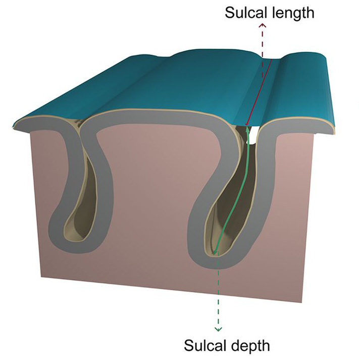
Courtesy of Jannsen et al, J. of Neuroscience
The cortical folding patterns in humans also change with age, establishing their final structure late in prenatal development while becoming less prominent in old age.
“While sulci can change over development, getting deeper or shallower and developing thinner or thicker gray matter — probably in ways that depend on experience — our particular configuration of sulci is a stable individual difference: their size, shape, location and even, for a few sulci, whether they’re present or absent,” said Bunge, who studies abstract reasoning in young people, from 6 years of age through young adulthood.
The smallest grooves, many of which are uniquely human, are called tertiary sulci because they appear last in prenatal development and are never as deep as the major or primary sulci that are most evident on the cerebral surface.
Scientists have speculated that the tertiary sulci emerge in parts of the human brain that have expanded the most throughout evolution and have a protracted development, and that they are likely associated with aspects of cognition — reasoning, decision-making, planning and self-control — that develop over a protracted adolescence.
But prior to this study, evidence was lacking for a connection between tertiary sulci and brain connectivity. The UC Berkeley study is one of few, all within the past few years, to provide such proof.
Sulci linked to cognition
Weiner and Bunge said that, as undergraduates, they were never taught how to define tertiary sulci; they often examined scans of average brains that did not match any specific individual.
Courtesy of pexels
Weiner noticed this mismatch as an undergraduate.
“At the time, all I knew was that I had some cortical squiggles that weren’t in the average brain atlases that we had in the lab. So the question I asked my mentors, Sabine Kastner and Charlie Gross, was: Do I have different structures that aren’t in our atlases or are structures missing from these atlases?” he said. “That sent me down a 15-year rabbit hole studying one particular tertiary sulcus in the visual cortex.”
That work showed that a specific sulcus, the mid-fusiform sulcus, varied in length from as small as 3 millimeters to as long as 7 centimeters in any given person. Moreover, the longer the sulcus, the better a person was at processing and recognizing human faces.
“About 2% of individuals have developmental prosopagnosia, which means they can’t perceive faces, and they don’t have any brain damage,” he said. “That sulcus, especially in the right hemisphere, is shorter and shallower in those folks than in what we refer to as neurotypical controls.”
Building on that rabbit hole, Bunge and Weiner wondered whether tertiary sulci in other regions of the brain, outside the visual processing units, also correlated with cognitive ability. Upon moving to UC Berkeley in 2018, Weiner began investigating the prefrontal cortex — located in the front of the brain behind the forehead — in collaboration with Bunge, who wanted to test whether sulci in this area would be linked to reasoning.
In a 2021 paper, the two collaborated to define all the smaller sulci in the lateral prefrontal cortex and created a computer model that identified the tertiary sulci as contributing the most variation in reasoning ability.
“The model identified that there’s tertiary sulci in the lateral prefrontal cortex that are contributing to reasoning skills in kids,” Weiner said.
Expanding on that work in the new study, Weiner, Bunge and their colleagues painstakingly catalogued the tertiary sulci in the lateral parietal cortex, located under and just behind the crown of the skull, and investigated its functional connections with the sulci of the lateral prefrontal cortex. For both studies, they studied 43 participants, 20 of them female, who ranged in age from 7 to 18. While in a functional magnetic resonance imaging (fMRI) scanner, the participants were given a reasoning task. The researchers focused on the brain activity in 21 sulci they had identified in each hemisphere of the brain, and the functional connections between these sulci — including, for the first time, tertiary sulci.
Across these individuals, greater depth for several of the sulci implicated in reasoning was associated with higher network centrality across the set of prefrontal and parietal sulci.
Experience affects sulci
Bunge pointed out that the association between depths of sulci and reasoning does not hold for all sulci, and that sulcal depth may change with experience.
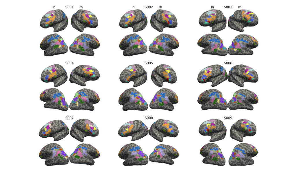
Courtesy of Hakkinen et al
“Do we think that an individual’s capacity for reasoning is set in stone based on their cortical folding? No!,” she said. “Cognitive function depends on variability in a variety of anatomical and functional features and, importantly, we know that experience, like quality of schooling, plays a powerful role in shaping an individual’s cognitive trajectory, and that it is malleable, even in adulthood.”
Weiner’s lab is creating a computer program to help researchers identify tertiary sulci in the human brain. Most programs only identify about 35 sulci, but when tertiary sulci are included, there are over 100, he said, including new ones that their labs have uncovered together. They argue that sulci could serve as landmarks to compare brains between individuals, since brains vary so much.
“Dozens of brain maps have been proposed in just the last five years, but they disagree about the areas of associated regions in the cortex, and there are mismatches between areas at the group and individual level,” Weiner said. “Examining network architecture based on individual sulcal morphology circumvents these disagreements and mismatches, with the opportunity to glean network-level insight from the local sulcal anatomy that is specific to a given individual.”
Aside from Bunge and Weiner, other UC Berkeley co-authors of the paper are former postdoctoral fellow Suvi Häkkinen, former graduate student Willa Voorhies, former undergraduates Ethan Willbrand and Jewelia Yao, and former visiting scholars Yi-Heng Tsai and Thomas Gagnant.
The work was supported by the National Institutes of Health through grants from the Eunice Kennedy Shriver National Institute of Child Health and Human Development (R21HD100858) and the National Institute of Mental Health (R01MH133637), and the National Science Foundation (CAREER Award 2042251).