Berkeley Talks: For Nobel laureate Randy Schekman, it all began with pond scum and a toy microscope
“I just could not believe the world that was revealed,” said the UC Berkeley professor of the microorganisms he saw through the plastic lens. He went on to win the Nobel Prize in Physiology or Medicine in 2013.
November 28, 2025
Follow Berkeley Talks, a Berkeley News podcast that features lectures and conversations at UC Berkeley. See all Berkeley Talks.
When UC Berkeley Professor Randy Schekman was 12, he scooped up a jar of pond scum and examined it under his toy microscope.
“I just could not believe the world that was revealed,” he said during a campus event earlier this month. “This complex set of creatures that you can’t see with your naked eye, and yet are moving and somehow mechanically independent, and able to do amazing things. And this was so fascinating.”
Schekman went on to become a professor of molecular and cell biology at Berkeley and win the Nobel Prize in Physiology or Medicine in 2013 for his discovery of how yeast membranes work. His research has led to advances in food and fuel production, as well as life-saving drugs and vaccines.
In this Berkeley Talks episode, Schekman explains the molecular building blocks that define who we are, the cellular processes that drive health and illness, and how curiosity-driven research leads to revolutionary insights into disease and opens doors to new possibilities for medicine and human health.
This lecture, which took place on Nov. 7, was sponsored by UC Berkeley’s Osher Lifelong Learning Institute.
Watch a video of Schekman’s talk.
(Music: “No One Is Perfect” by HoliznaCC0)
Anne Brice (intro): This is Berkeley Talks, a UC Berkeley News podcast from the Office of Communications and Public Affairs that features lectures and conversations at Berkeley. You can follow Berkeley Talks wherever you listen to your podcasts. We’re also on YouTube @BerkeleyNews. New episodes come out every other Friday. You can find all of our podcast episodes, with transcripts and photos, on UC Berkeley News at news.berkeley.edu/podcasts.
(Music fades out)
Matt Shears: Good morning, OLLI@Berkeley, and welcome to our speaker series. We are happy to have you. As many of you know, we ended our fall term courses yesterday, and so we would like to point out that if you would please fill out your evals for your courses. We use those to help guide our programming and to help our instructors receive feedback in a constructive way.
And if you aren’t quite ready to end your term, we do have one further event. It’s the SF Opera Primer with Carol Christ and Nanette Coleman, and they’re going to be talking about The Monkey King next Tuesday, Nov. 10. You can find information about that on our website. But for today’s main event.
Randy Schekman is a Nobel Laureate and professor of molecular and cell biology at UC Berkeley. He’s the former editor-in-chief of Proceedings of the National Academy of Sciences and the former editor of the Annual Review of Cell and Developmental Biology. His research and work has helped reshape our understanding of life itself. Today’s talk is “Genes, cells and discovery in basic science and disease.” Randy, welcome.
Randy Schekman: Thank you very much. Pleased to join you today, and to tell you a little bit about myself and how I got into the research that I’ll describe to you. I’m a creature of the University of California. I was an undergraduate at UCLA from 1966 to 70. I had four years of misspent youth as a graduate student at Stanford before I then have spent the rest of my life at the UC system. Then as a postdoctoral fellow at UC San Diego, and now beginning in 1976, I’m now in my 50th year on the faculty at Berkeley. So I’m part of the woodwork here.
Well, I grew up in Southern California. And for reasons that I can’t really recreate, I developed a very early interest in microorganisms. I had a toy microscope when I was maybe 12 years old. And I remember going to a local river and it’s a pond of still water.
And I collected a jar of the scum on the top of the pond. And I brought it home. And I looked into this plastic lens of my toy microscope and I just could not believe the world that was revealed. This complex set of creatures that you can’t see with your naked eye and yet are moving and somehow mechanically independent and able to do amazing things. And this was so fascinating.
I attempted to recreate this for my parents and brothers and sister at dinner one evening. And my father, who was an engineer, was really very skeptical. He thought, I’m sure, that I had a vivid imagination. And I was a little offended by his remarks. So I decided to save my earnings. I used to babysit and mow lawns and deliver newspapers. And I was going to build up the princely sum of $100 in 1962.
So I put the money in an envelope in my bedroom. But my mother, I couldn’t quite get the $100 that I needed because my mother was borrowing the money. So one Saturday morning I finished mowing a neighbor’s lawn. And I was so upset. I rode my bicycle to a local police station and I told the duty officer that I was running away from home because my mother was stealing my money.
And so they called my father into the police station and he went and talked to the captain. And he looked and came out looking rather upset. But we went to a local pawn shop and bought for that $100 this microscope, which I used for the rest of my time in school every year, mounting a science fair project, which really was the key to my interest in science. And in fact, on the bulletin board above my senior year was my letter of admission to UCLA.
Although initially a pre-medical student, I realized that science was my objective. And so I trained working in a laboratory there and then as a Ph.D. student at Stanford. And I learned the art and craft of studying complex biological processes by taking them apart piece by piece. So let me tell you what we’ve been doing for the past 50 years at Berkeley.
So this is an image some of you may have seen. It’s a representation of chromosomes from a cell that’s dividing in your blood, and we can visualize all the diploid, two copies of each chromosome, all 23 of them, with fluorescent molecules that probe the different DNA sequences in the chromosomes. I use this to illustrate the complexity of the human genome. All these chromosomes are housed in the nucleus of a cell. And they are the blueprint for 23,000 different genes which code for approximately 100,000 different protein molecules.
All those molecules are made inside of cells and they catalyze the chemistry of life, or they form structures that give rise to cells that are able to nourish themselves and grow and divide. Now, what has been a major interest in the area of cell biology is how these cells organize themselves into compartments and how some of these compartments contribute to a process of protein export from cells. Now, although all those 100,000 proteins are made inside of the cell, maybe 20% of them or so are shipped outside of the cell by an elaborate export machinery that I’ll describe.
This machinery evolved on earth probably over 2 billion years ago in microorganisms and has persisted to this day to allow us to organize, for instance, the secretion of molecules like insulin or blood proteins or growth factors or even the chemical neurotransmitters that mediate communication in the brain.
Now all cells are surrounded by a membrane. This is the simplest cell in our body. These are the red blood cells that carry oxygen bound to hemoglobin to the tissues in our body. Membranes have a quite interesting structure. They are basically two leaflets of phospholipid molecules. These red molecules are lipid molecules that have a hydrophilic polar water-loving surface facing the outside of the cell or the inside of the cell. And they have these long strings of hydrocarbons called fatty acids that are very hydrophobic. They don’t like water. So they form the interior of a membrane bilayer.
Now, these lipids by themselves, if they were the only things in a membrane would be insulating, they wouldn’t let anything in and out. Polar molecules, like ions or sugars, can’t go through a pure lipid bilayer. So evolution has built protein molecules into membranes that give each membrane a different personality.
Some of these proteins are channels through which small ions or small molecules can pass or be concentrated in the cell. Other molecules are receptors, like the receptors for hormones such as insulin. The interesting thing about membranes that fascinated me for my independent career was that unlike protein molecules or nucleic acids, which are covalent structures that don’t change over a short time course, membranes are fluids. They’re two-dimensional fluids, because the lipids are in a fluid state in living cells. That means that proteins and lipids are free to move about laterally or to spin about an axis perpendicular to the membrane.
So thinking about how a membrane gets put together is a really different kind of problem than thinking about how one can put together proteins in nucleic acids. Now, here’s an example of one of the most complicated cells that’s specialized in the secretion of insulin.
This is the beta cell in our pancreas. In between meals, insulin is made inside the cytoplasm and then becomes encapsulated within membranes called granules. And these granules house what you see here, a crystalline form of insulin. This builds up in between meals. And then when you have a meal and you start to adjust polysaccharides, carbohydrates, sugars like glucose get released, coarse through your body, and then these granules move up to the perimeter of the cell and discharge their content.
As you’ll see in the next slide, oops, you don’t see it here. They discharge their content by a process of membrane fusion where the granule membrane merges with the membrane surrounding the cell. And the crystalline form of insulin is discharged to the outside of the cell. It dissolves, courses through your body, goes to the various tissues, and those tissues that bind insulin at a receptor swallow up all the glucose and store it for a time being into a molecule of call it glycogen, which can be used as a storage form of sugars to used later for energy production.
Now this process of capture of molecules for discharge at the cell surface is repeated all over your body. Here’s an example of such a situation where instead of granules housing insulin, these little membranes called vesicles at a nerve terminal, specifically called synaptic vesicles, carry chemicals that transmit information between cells. In this case, a nerve cell is communicating with the muscle cell. Or in the brain a nerve cell is communicating with billions of other nerve cells.
Now, this is exquisitely controlled by these synaptic vesicles, touching docking on the inside of the nerve terminal. And when these nerve cells are activated at a key moment, there is an explosive membrane merger, membrane fusion that connects the vesicle to the plasma membrane, causing chemical neurotransmitters to be secreted in this case into the space, the synapse between a nerve cell and a muscle cell.
These chemical neurotransmitters, molecules like dopamine or serotonin or acetylcholine, bind to receptors on the other side of the cell, in this case a muscle cell, and cause the muscle cell to contract. But in the brain, these networks are purely nerve cells touching each other. And they make sometimes it’s calculated to be a hundred trillion different connections in our brain. And every aspect of our personality, our memory, all of our abilities, our physical and mental abilities, are controlled by a form of communication that ultimately concludes in a very rapid and controlled discharge of chemical neurotransmitters secreted when these vesicles fuse, merge with the plasma membrane and dump their contents to be exposed to an adjoining cell.
Now in the mid-1970s, some really great cell biologists used tools such as microscopes, such as the images that you’ve seen a moment ago, the electron microscope, to look inside of cells. And one of the great founders of the field of cell biology was a Romanian scientist who did his work in New York at the Rockefeller University.
In 1974, George Pallotti won the Nobel Prize for figuring out how molecules like insulin are encapsulated in stages as they move within the cell through a series of membrane-bound compartments. They start being made by ribosomes, which stitch together amino acids, one after another to make protein molecules and molecules like insulin then are threaded across a membrane into the salmon-colored interior, a a canal network of membranes referred to as the ER. From there, some molecules become packaged into small membranes, small vesicles, and convey their content to a structure that was first described in the 19th century by an Italian neuroscientist by the name of Camillo Golgi.
It’s now called the Golgi apparatus in honor of his discovery. And it took more than 50 years until Pallotti was able to show that the Golgi apparatus is a bus stop in the course of transit of molecules like insulin from where they’re made to the cell surface by this membrane fusion process.
Now, what was most interesting to me when I was considering my independent career was it was known that these membranes communicate by this pathway. And it was known to be of general importance in all cells that have a nucleus so-called eukaryotic cells. But in 1974, when Pallotti won his Nobel Prize, absolutely nothing was known about how this works. What are the nuts and bolts, what is the mechanism that allows these molecules to progress in this very complicated dance within the cell? And so what I decided to do in 1976 when I began my career at UC Berkeley, was to explore this process in the microorganism.
Again, my favorite experimental subject had always been to study microorganisms. And I chose to work on a really simple microorganism baker’s yeast. You know baker’s yeast because of course, it’s so important production of alcohol and in baking bread, it’s been industrialized for thousands of years.
But what you may not know is the yeast cells are also a really simple and powerful tool that biologists use to study complex processes that are characteristic of all cells that have a nucleus. So here’s an image, a special electron microscope image of a yeast cell that’s caught in the act of growing and dividing in a rich medium nutrient broth. The cell starts as what is called the mother cell. And if the nutritional conditions are good, these cells start to grow and divide. And they do so not by dividing in half, but rather by sending a small bud that emerges from a particular point on the surface of the mother cell.
And during the 90 minutes or so that it takes this bud to enlarge, it eventually becomes the size of the mother cell as you see on the left. Now, we reasoned in my lab that new surface material had to be added to this bud, and perhaps new membrane had to be added just underneath this thick cell wall to achieve the size of the mother cell, at which point the daughter cell divides, separates from the mother cell. And if yeast cells are growing in a good nutrient broth, they will continue to grow exponentially producing billions and billions of identical copies.
Now, one of the virtues of using yeast is you can grow lots of it cheaply, you can break the cells open and isolate molecules, but you can also apply a very simple genetic tool that is used typically in microorganisms, but which is much more difficult in diploid organisms like us.
If you expose a yeast cell, which grows in what is referred to as a haploid life cycle where only one copy each of the 6,000 different genes is present in a cell. If you expose such a cell to a chemical that causes mutations randomly, you will sometimes introduce a change into a protein that makes it, for instance, unstable. Unstable in a way that allows the cell to grow If you grow the cells at room temperature, but unstable if you warm the cells up to a higher temperature such as human body temperature, that range of temperatures is perfectly adequate for a normal yeast cell.
But if a cell is crippled in a very special way by a mutation in an otherwise essential protein, the cell will form a colony on a Petri plate at room temperature. But that colony will not form another colony if the plate is copied onto something that’s incubated at human body temperature.
Those are called temperature-sensitive lethal mutations, and they’re very easy to obtain and extraordinarily useful in evaluating the function of genes in a simple organism like yeast. So I had the good fortune not only of a good simple idea, but a great graduate student shown here in 1978. One of my first graduate students, his name is Peter Novick, and he was masterful in the laboratory. And the two of us then began a search for mutations that specifically cripple the process of protein secretion.
That is how molecules that yeast need to grow, for instance, on the surface of a grape need to be exported outside of the cell to scavenge macromolecules from the environment so that yeast cells can be supplied with adequate nutrients. Peter was enormously successful, so much so that later in his career in his own independent laboratory, he has now become the George Pallotti chair of cell biology at the University of California San Diego.
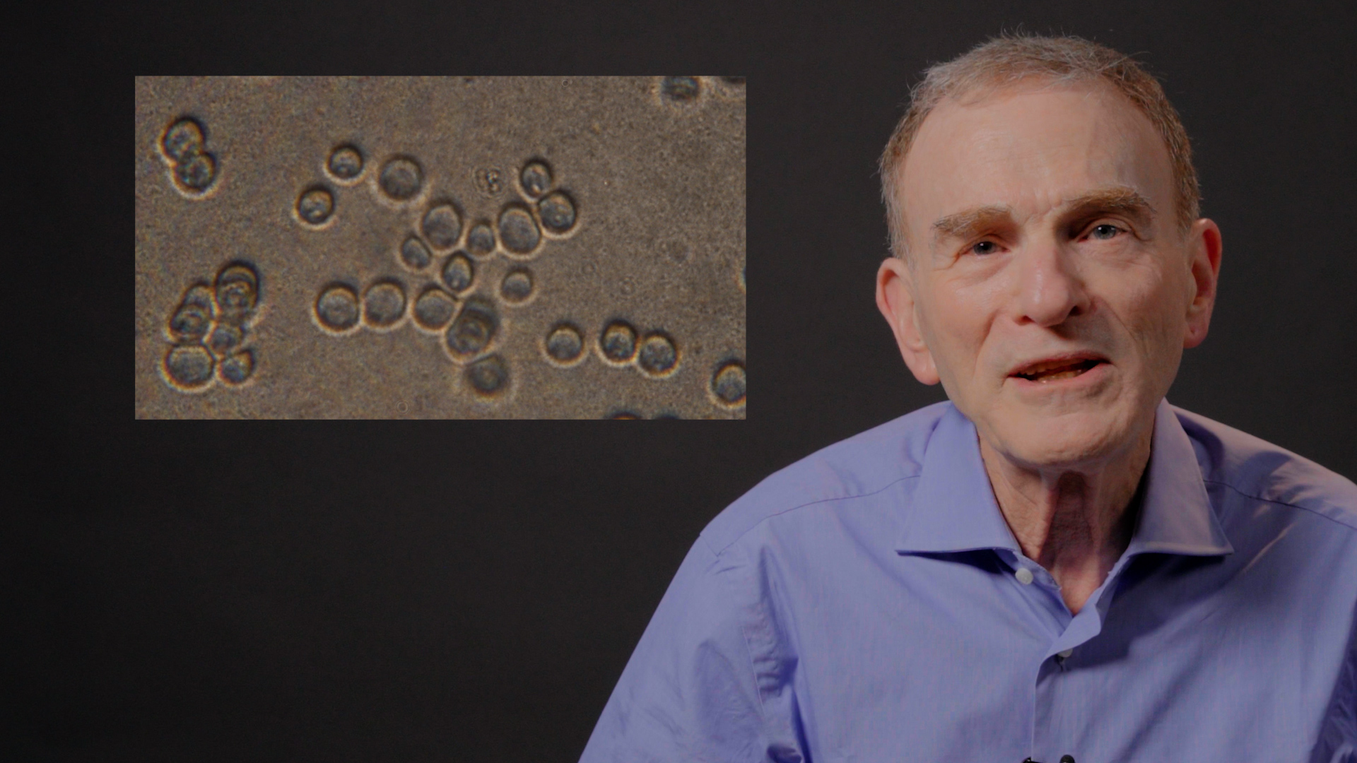
So here’s another image of a yeast cell that we knew of in the literature in which we were able to obtain ourselves. The cell is much less dramatic than the beta cell of the pancreas. But still there’s a lot going on. Most of it is going on because these little granules that you see in the cytoplasm are the machines ribosomes that stitch amino acids one next to another to make the 6,000 different proteins that yeast cells need to grow and divide. Yeast cells of course, also need to secrete, to export some molecules to be able to gather things in from the environment.
Now, our attention, Peter’s and my attention, was drawn to these small membranes. They look like those synaptic vesicles that I showed you earlier at a nerve terminal. But of course, yeast cells aren’t communicating with neurotransmitters. They are conveying enzymes that are made inside the cell to the outside of the cell where they can scavenge macromolecules from the environment.
Now, Peter and I thought in addition that the membrane bilayer that surrounds each of these vesicles could be a building block for the growth of the plasma membrane, the membrane that surrounds the cell. And so we guessed that every time a vesicle merged by fusion, not only would things be secreted to the outside of the cell, but the membrane would become a building block for the stepwise growth of the bud. Thus, this process of traffic of vesicles and membrane fusion would be essential to allow the bud to get bigger in order to promote cell division. So that of course, predicts that the genes required for this process, for formation of vesicles, for their migration and fusion would be essential.
And thus, it would be useful to look for those kinds of mutations that I described a moment ago that are temperature-sensitive lethal. So one of the great moments in my career came when Peter, after having isolated the first such mutant called SEC1, short for secretion, defect number one, when he took these cells and incubated the cells in a culture at a higher temperature, human body temperature for a couple of hours, he looked at these cells by microscopy and he called me down to the microscope room in the basement of Barker Hall very excitedly to show me the following picture.
Instead of the small number of vesicles that are found in a normal bud involved in growing the cell, these cells fail in that last step because they have a mutation that cripples a protein that’s required for the membrane to merge by fusion. And instead, the vesicles now build up in the thousands, spilling over and filling the entire cytoplasmic volume, so much so that the cell eventually dies of a kind of molecular constipation because it can’t get any bigger, and yet it’s making all this stuff. And yet before the cells die, if you return these cells back down to room temperature, the vesicles can reengage and be trafficked and protein secreted, indicating that that mutation caused a thermally reversible defect in the protein product of the SEC1 gene.
So we then were very excited by this and refined our procedures, and we’re able to isolate hundreds more such mutations in an effort to define the entire pathway that yeast cells use to see if it’s similar to what human cells use.
And so after the next few years and further brilliant work by other students, we were able to populate the entire pathway with a whole bunch of genes. In addition to Sec-I, there were nine other genes required at this last step of the pathway. At the very beginning of the pathway, we found genes that code for a channel in the ER membrane, through which polypeptides must pass to begin the process.
So lots of genes formally looking very much like the pathway that Pallotti and his students had recreated for the pancreatic secretory cells. But importantly, with each step informed by a number of genes and thus a very specific number of protein molecules. Now, after we assembled this map, we began to use then the emerging techniques of molecular biology to obtain the genes for each of these relevant mutations to the corresponding normal gene. And when we sequenced the DNA of each gene, we found a bunch of genes that didn’t look like much of anything then in the genome databases.
But within a few years, the human genome was sequenced, and we found, perhaps not surprisingly, that the very genes that we had obtained and discovered in yeast have their counterpart in the human genome. In fact, this gene, SEC1, the first gene has its equivalent in the brain. And that gene, that protein in humans is responsible for allowing those synaptic vesicles to dock in preparation for fusion and neurotransmission. And in fact, all of the genes are similar.
Now, this of course illustrates a point that I made earlier, and I emphasize the point of conservation of evolutionary processes as depicted here by a cluster of yeast cells, such as you might see growing on the surface of a grape. And the great evolutionary biologist, Charles Darwin. And I submit to you that the machinery inside of yeast cell, the lowly yeast cell, evolved 2 billion years ago on Earth.
The machinery that blueprint, those sets of genes required to do things inside of cells are conserved in humans, certainly in Charles Darwin. So the Charles Darwin brain as elegant and exquisite as is relies on machinery that evolved on Earth 2 billion years ago. Now, one interesting application of this, which I had not anticipated, was that when we made these discoveries in the San Francisco Bay Area, the biotechnology industry had just emerged. And I helped a local company called Chiron to engineer yeast cells to manufacture large quantities of clinically useful human proteins.
For example, once the gene for human insulin had been obtained, it was possible to engineer that gene into yeast cells and to allow yeast cells to churn out what almost certainly is metric tons of insulin into the growth medium. And now many years later, 30% of the world’s supply of human recombinant insulin used to treat diabetics is grown in giant fermentation vats, tens of thousands of gallons large, churning out yeast cells that are spitting out insulin using the yeast secretory pathway.
The same was true with this company when it became important in the 1980s to have a new source of a vaccine against hepatitis B. The hepatitis B viral surface protein is a membrane protein, and they found, this company found, that if you put that gene into yeast cells, the viral antigen, that’s the basis of the vaccine is assembled into little vesicles.
And so for anybody here who may have had the Hep B vaccine, in fact, most newborn children are vaccinated very quickly. That vaccine, those injections were little vesicles made in yeast cells, housing just that protein and not the infectious virus. Now, I was trained as a reductionist biochemist. So the genetics, the mutants, and all these genes were of course a useful tool in my mind to understand really the nuts and bolts. What do these proteins do that are coated by these genes?
What I’ve said so far is that it’s conserved that we have a blueprint, but the blueprint can’t be read unless you understand what the protein molecules that are coated by these genes do. So for that purpose, it was essential in my mind to have a way of isolating these molecules and studying their function in the test tube. And along the way, other people in the lab were able to show that this first step in the pathway that we had seen could actually be broken down into two steps.
That is when yeast proteins are made and assembled in the ER, they get packaged into small little vesicles. And these little vesicles then merge by fusion at this structure that I told you about called the Golgi apparatus. Now, this refinement in our discovery based just on use of genetics and microscopy, allowed us to devise a biochemical process using broken preparations of yeast cells to manufacture these little vesicles by a process of membrane budding from the ER.
This could be studied in the test tube, and we could show that this part of the pathway was directed by the concerted action of a set of proteins, largely cytoplasmic soluble proteins that pinch a vesicle by budding from the ER. And we purified the proteins that are responsible for this pinching, budding mechanism. And when these pure proteins that we had obtained are mixed with isolated ER membranes in the test tube, they produce a uniform population of small coated vesicles. You see, they have a fuzzy coat on the outside. We call these vesicles COPII vesicles, and we and others have shown in the years since then that the COPII proteins are not only responsible for pinching the vesicle, but by directly capturing membrane proteins, they can actually gather in those molecules that are designed to be moved from the ER of the Golgi apparatus. And this is true in all eukaryotic cells.
Here’s a higher magnification view of these coated COPII vesicles where the fuzzy coat are the actual molecules that we purified to reproduce this reaction. Now, crystallographers who study protein structures then took what we had discovered and were able to obtain a high resolution image at the atomic level of how this coat actually envelops and pinches a membrane.
And I show this just as a beautiful picture that the coat, this COPII coat, has two parts, an inner layer that touches the cytoplasmic surface of the ER membrane and grabs onto membrane proteins that are destined to be moved along. And that inner coat then is surrounded by an interesting polyhedral network of two other SEC proteins that form a unique so-called cube octahedral structure that surrounds the membrane, shapes it into a small vesicle, and then separates it by a fission, membrane separation fission, to make a transiently coated vesicle.
Now, I tell you about this because there’s a really interesting story that emerged after we made our discoveries. And started to explore the same process in human cells, much the same mechanism operates in human cells with some interesting differences.
Subsequent to our work on this in human cells, I had a chance exchange with a clinician then at Johns Hopkins University, a scientist named Simeon Boygev, who had in turn collaborated with a colleague, a clinician in Riyadh, Saudi Arabia. They had found a Bedouin family that had a rare craniofacial disorder. There are many genetic disorders that affect the shape of the face and the head, but this one was different than the others, and it seemed to affect the maturation of the skull and about that the changes in the shape of the face shown here in images of two children who carry two bad copies of a particular gene.
These kids, for instance, the soft spot in the top of their head never closes, and it’s because of a failure in skull morphogenesis, bone morphogenesis. They develop cataracts at an early age. They have very brittle bones, but surprisingly, in spite of these mutations, the kids survive and they’re fragile, but they’ve survived.
Now, what was of course interesting to us was that the human genetics showed that the mutation in these kids is in one of the genes that we discovered in yeast, a particular gene called SEC23. In humans, this gene has two copies. In yeast, it has only one copy, and those two copies are expressed, are represented in, different tissues in the body. And in this case, one particular copy, the one that had a mutation, had a change in one amino acid of the protein that is absolutely conserved in all of the molecules of that gene found in nature.
So a change in essential amino acid caused that protein to malfunction in these kids in the tissues in which that particular copy of the gene is represented. Now, we obtained from the clinician in Saudi Arabia, samples of skin from one of the parents. The parents are perfectly normal. They are genetic carriers. The mutation is said to be recessive. That is one copy is sufficient for normal growth, but if you have two bad copies, you see this defect.
So we had a sample of a normal skin cell, and then a sample from one of the parents, and then from one of the kids. And we had it looked by cutting thin slices through these skin cells, and the results were really very dramatic. The top image is a thin slice through a normal skin cell, and you see these ER membranes with some kind of granular material inside in what is referred to as the lumen, the canal-like network that I started this presentation with.
In the middle panel, it’s a slice through one of the parents and the ER membrane is beginning to look a little misshapen. And some stuff is accumulated, but it’s not enough to cause any pathology. And yet when the cells of one of the kids are grown in the laboratory, the ER membrane is completely distorted.
And we showed in experiments in the laboratory that these cells have a great deal of difficulty trafficking molecules from the ER, the Golgi. They pile up in the ER. One of the major molecules that we’ve shown to accumulate in the ER in these cells is collagen. Collagen made by skin cells, collagen made by connective tissue. This seems to have a particular bottleneck in the capture of molecules because of a defect in one of the proteins in the COPII coat. OK? So we were having a lot of fun exploring basic science and seeing it applied by the biotechnology industry.
But at a certain point in 2013, my life changed quite dramatically. I got a call at 1:20 in the morning from Stockholm informing me that I had won the Nobel Prize. And from that moment on, my life changed quite considerably. Two months later, my family joined me in the celebration. We went to Stockholm, where I was greeted by the king to receive my medal. But earlier than that, something quite unexpected happened.
And that is every year the new laureates are given the opportunity to provide some artifacts from their laboratory to be put on display in what is called the Nobel Museum in Stockholm. Well, I didn’t think that Petri plates or toothpicks would be particularly interesting or novel, but it turns out that old microscope that I had that was my pride and joy throughout high school, had been saved by my parents. They sent it up to me when I had my family here in Berkeley.
Neither of my children, regrettably, ever had any particular interest in science. So the microscope sat around and collected dust until I got that email from Stockholm. And I dusted it off and I sent it to Stockholm. And if you ever go to Stockholm, you can go and see my microscope here on display in the Nobel Museum together with a caption in Swedish and in English, saying how I at the age of 12 had to run away from home to begin my career in science. So thank you very much for your attention, and I’m now would be happy to answer any questions that you may have.
Matt Shears: Thank you very much, Randy. Folks, if you do have questions, you can put them into the chat and we will get them to Randy that way. Maybe as we wait, Randy, why don’t you tell us a little bit more about your trip to Stockholm in December of 2013. What was your biggest surprise in joining that illustrious group?
Randy Schekman: Well, it was in some ways complicated. And there’s a story that I can tell while we’re getting some questions. Although my life has generally been blessed with many great moments. I had a serious problem when my wife was diagnosed with Parkinson’s disease at age 48. And so over the next 20 years, she progressed, it became more severe. She developed dementia in 2012 and was required pretty much full-time care, including during the trip to Stockholm.
So fortunately, my family was with me, and they were able to help with that, and she was still sufficiently conscious to appreciate what was going on. And so I’m grateful that she and my 86-year-old father were there to join a celebration, but it got much worse. And in almost four years later, she died in the middle of the night for reasons that were never really entirely understood.
So life comes with its challenges, and that was certainly one of them. But the time there was a joyous event, it’s exhausting. As soon as you land in Stockholm, they meet you, not at the gate, but as you leave the fuselage of the plane, they’re there just in the gangway and they take you down the steps to a limousine to take you to the VIP room. And then from then on, they control every movement. It’s exhausting. Ten days of exhaustion. The place is full of viruses. So I caught a cold after three days, and it was even more exhausted, but generally quite a remarkable experience.
Matt Shears: Yeah. I’m glad that your dad and your wife were able to experience it with you. It looks like we do have some questions coming in. Here’s one. How do you see AI, the impact of AI … Let me rephrase. How do you see AI impact the rate of studies in your field and potential new discoveries?
Randy Schekman: Yeah, it’s a revolutionary tool. I don’t view it as a threat as some people do, but as an incredibly powerful tool. And one example that some of you may be familiar with is the now ability using AI to predict what proteins will look like when they fold into compact three-dimensional structures.
So it’s been possible experimentally to determine atomic structures of proteins using powerful tools of crystallography and electron microscopy. Tens of thousands of such proteins had their structures solved experimentally. But four or five years ago with an algorithm developed by a company called DeepMind in London, program called AlphaFold, they were able to predict the structures of every molecule that has ever been predicted to exist on Earth.
And they deposited online a library of 250 million different structures, all because of AI. Many of them are subsequently been confirmed experimentally. It’s an incredible tool for those of us who aren’t crystallographers to get the structure, but to have the structure of a protein that we’re working on. Enormous tool. So that’s just a taste of what AI will deliver.
Matt Shears: Wow. April says there’s been huge progress learning about cell processes during your career. What do you think is the next scientific frontier for molecular biology?
Randy Schekman: Well, first of all, when you win an Nobel Prize, unfortunately they don’t give you a crystal ball. So I am just speculating. But I think most of us would agree that the next century will be the century of the brain, figuring out all those connections. It’s just vast. Think about this, AI is incredibly powerful and the computational power, predictive power of AI will exceed that of the human brain. But there’s enormous difference in scope.
In AI, some of these companies are now having to recommission nuclear power plants to generate enough energy to drive these vast farms of servers. But we rely on the energy of about a 20 watt light bulb contained within our skull to do things that AI can’t yet do, to be creative, to use our imagination. So the brain is a mystery. As an example, one goal eventually would be to figure out the wiring diagram of the entire brain.
With 10 billion cells, the wiring diagram is not yet possible to deduce. In fact, this year, for the first time, it’s been possible to deduce the wiring diagram of the fruit fly brain, that little thing. It took all this time to work out the wiring diagram of that little brain. This year also, it was possible to work out the wiring diagram of one cubic millimeter of a mouse brain.
And so doing the whole mouse is going to take a lot more, and doing the human brain, it’s going to take a leap of technology. So just the wiring diagram, let alone understanding the flow of information in the brain, that is going to keep us busy for a century or more. And so I think a lot of young scientists see that opportunity and use their creative instinct to try to think of new ways to plumb the mysteries of the brain.
Matt Shears: That’s amazing. Richard asks, are there implications from your work to human reproduction?
Randy Schekman: Yes, of course. So cells of the egg and then the fertilized early embryo, as they divide, one of the interesting things that we’re working on that is very relevant to very early embryonic development is at one of the earliest stages, the embryo that’s growing is not yet implanted on the uterine wall. And when it implants after two weeks after fertilization, that gives rise to the placenta, which is essential to nourish the embryo as it develops.
In the outer layer of the pre-implantation embryo as a layer of cells called trophoblasts. And at a key moment, the cells in that layer fuse by this process of membrane fusion that I talked about in earlier. It’s a very different kind of fusion process that produces a layer basically of cytoplasm that surrounds the embryo. And then the embryo attaches to the uterine wall, and that gives rise to the placenta.
Now, we’ve been studying the membrane protein in those trophoblast cells that promotes this merger of all these cells in a layer to create what is referred to as a syncytium. And we’re very interested in that fusion reaction as it relates to the communication of other cells in the body, because it turns out that gene, the gene for this molecule that makes fusion of the trophoblasts, is usually only expressed in the early embryo. It’s a gene that was borrowed, if you will, during evolution by an ancient retroviral infection. And that virus has used this protein, Evolution is used that protein to promote this fusion reaction.
Now, interestingly, and this is relevant to work that we’re doing, interestingly, tumors, cancer cells turn on that gene. And we think that gene now in tumor cells may be responsible for tumor growth and possibly metastasis. So we have some projects in the lab that are working on that. They don’t necessarily relate directly to the earliest stages of embryonic development, but some consequence of the genes that are required for that process. So yes, there’s a connection
Matt Shears: Different Richard. Richard asks, what prevents the target genes from mutating in yeast within the large vats producing insulin or other proteins?
Randy Schekman: That’s a great question. Of course, this goes on all the time. And so every time you start a new yeast culture, whether it’s on a small scale or on a big scale, you have to make sure that the cells are growing normally as they normally do. But you can’t prevent these mutations from happening. But it turns out that most mutations are silent. That is they don’t really affect the cell that much. They don’t change enough to be of any concern, but a cell will occasionally get a mutation that’s crippling. And so you have to make sure that the seed stock that you’re using to grow a new culture behaves normally, at least on the Petri plate.
Matt Shears: And Ken wonders, how do you think CRISPR technology will impact the future of your research?
Randy Schekman: Well, it’s not the future, it’s the present. Everybody uses it as a tool now. It’s the tool that has allowed us to do in human cells growing in the laboratory what we always could do with microorganisms. That is to introduce mutations or to remove genes to manipulate a cell, even when it’s a diploid cell with two copies of each gene.
So it’s an enormously important experimental tool, and not to take away at all from its practical applications. We now, in the past two years, have two examples of the use of CRISPR technology to cure human genetic diseases. First was the ability to repair a defect in patients with sickle cell anemia. This is a mutation that was first recognized by the great 20th century chemist, Linus Pauling, to be in the gene that codes for one of the subunits of the oxygen carrying protein hemoglobin.
It’s now possible to replace that mutant protein with a normal one in young patients who suffer from sickle cell anemia. And several patients have now fully recovered and are normal functioning young adults because of this, at the advent of CRISPR technology. Even more recently, what would’ve been an invariably fatal disease was cured in a young kid just within a few months of birth. A mutation that would’ve killed a kid by the end of the first year, they rescued him by changing that mutation back to normal with CRISPR technology. So we’re going to have a library of CRISPR cures for all kinds of genetic diseases going forward. Enormously important.
Matt Shears: This is a bit of a follow-up. This is Noel wondering if you’re interested in rare disorders or if funding is limited so that there aren’t resources to study those.
Randy Schekman: Rare disorders, at least in the NIH before this year, had special funding sources through the NIH. And some companies have developed therapies based on rare disorders because of a privileged position they have in having their work supplemented by federal or private funds.
So there are lots of rare mutations. And patient groups focus on those particular types, and they lobby and have, at least in the past, been quite successful in getting additional funding for them. I have, as a result of my wife’s illness, I’ve had an opportunity to lead a large international organization focused on collaborative studies on the basis of Parkinson’s disease funded by the Sergey Brin Family Foundation. Mr. Brin has put down a billion dollars to our effort around the world to bring groups together to try to understand the basis of that disease. And so as a result of my wife’s death, I’ve taken this on now as outside my research, quite important activity.
So there’s a lot of money, both public and private and supportive science. But of course, it will be no surprise to people listening in that we are in peril in this country now because of a general lack of appreciation for science and for the scientific enterprise. And a concerted attack on the knowledge that we’ve accumulated because of basically the inappropriate choice of RFK Jr. to be the head of HHS. He is neither a physician nor a scientist, and he has some crackpot ideas about things like vaccines. And this now represents a clear and present danger for public health in America as a result of that.
Matt Shears: Yeah. Two questions from Martha. The first following up on Parkinson’s, what do you see as the major causes, or can you speculate based on what you’ve learned?
Randy Schekman: Sure. So we know that there are genetic influences. Mr. Brin shares a mutation with his mother in one of the 20 or so genes that are found in familial forms of Parkinson’s. His mother is quite ill, he’s at risk, but so far healthy.
So it’s genetics, but that’s only a fraction, maybe 20% of what we know to be. Most likely, Parkinson’s is largely, and probably in the case of my wife, the result of exposure to environmental toxins. They’re known pesticides that increase the incidence of Parkinson’s among people who use these pesticides in their career. And exposure to these even casually can send some people down a path that leads inexorably to Parkinson’s.
Part of what we’re doing in my study group called Aligning Science across Parkinson’s, ASAP, is to focus in on the molecules in pesticides to understand exactly what they do to cause damage that can be propagated in the form a progression of this disease. Because if we can understand how these molecules work, we can probably think of ways to counteract their action to cause this damage.
Matt Shears: And a follow-up from Martha, brain cells have a limited lifespan. How do our new brain cells re-encode the info from the dying cells?
Randy Schekman: Well, most of the cells in the brain do manage to live through our lifetime, but the neurons that are particularly vulnerable in Parkinson’s, they’re called dopaminergic neurons. They’re enormously complicated cells. They’re always on. They’re always firing. And they are particularly vulnerable both to mutations that I mentioned and to environmental toxins. So those cells do die. These dopaminergic neurons are dying as we speak in all of us. They tend to die at a greater rate in Parkinson patients for various reasons, but most nerve cells have effective pathways of repairing themselves.
There’s a very active pathway to patch up damage on the cell surface. Damage to one of the organelles that’s essential in all of our cells called the mitochondria. And it’s the powerhouse of each cell. Cells have evolved, over 2 billion years, pathways to get rid of damaged mitochondria and allow those that remain and that are healthy to take over. So cells are generally very resilient for the most part. And in their brain, they last for a lifetime. Some of them are replaced by a reservoir of stem cells, but most of them function for quite a long time and are quite resilient.
Matt Shears: Dorothea was wondering something along these lines. She asks, “What new projects are molecular biologists working on for autosomal recessive disorders like mitochondrial syndrome disorders?”
Randy Schekman: Yeah, yeah. Well, those are really challenging. The techniques of CRISPR are well-designed to work on nuclear genes, but mutations in the mitochondria have their own little genome, and there are some fairly, fortunately, rare genetic disorders of the mitochondrial genome. And getting CRISPR tools into the mitochondria to repair those mutations is more of a challenge. But there are approaches to doing this, but they’re not yet ready for prime time. So those are tough diseases.
Matt Shears: We do have a couple of comments in here. People expressing sympathy or empathy with respect to funding and the sciences, and the emergency that we are undergoing. April asks a question about this. Given the attack on science funding by the current administration, do you think that funding from other countries or private sources will blunt the decline?
Randy Schekman: Well, first of all, although it looked like it was going to be a disaster this year, fortunately, the troops have mounted an information campaign with Congress. And fortunately, both the House and the Senate have approved full support for the NIH, at least full support based on last year’s budget. And so piece by piece, it’s slowly coming back.
But it’s still a threat because Trump absolutely, he’s captured this atmosphere of antagonism to science. In his first administration every year he proposed a huge cut in the NIH budget, and every year he was overruled by Congress. We have a different kind of Congress in place now. And the hope is that we will have another different kind of Congress in place after next year. But those of us who are in the trenches are fighting, but the damage is already done. So many staff in federal agencies have been fired or quit.
Who in their right mind would want to take a job now in the federal government? And so these are people who are essential to doling out the money that’s been allocated. And so the money is coming, but very slowly. It’s trickling in. And there’s no replacement for philanthropy. We desperately need more resources in the University of California, the private institutions typically raise a lot more money than we do.
But Berkeley is getting much better at it. We now routinely raise over a billion dollars a year in philanthropic support. Our budget from philanthropy exceeds that given to us by the state. And that’s because we are no longer a public institution. We’re a publicly assisted institution.
Matt Shears: Well, let us hope that sanity returns to all of the approaches to science here. Randy, I don’t see any more questions in the chat. I’d like to thank you for joining us. We are pretty much at 11:30 a.m. If we can give Randy a virtual round of applause. Thanks for spending time with us today.
Randy Schekman: My pleasure. Thanks for the opportunity.
Matt Shears: Have a great afternoon everybody.
Randy Schekman: Thank you. Bye.
(Music: “No One Is Perfect” by HoliznaCC0)
Anne Brice (outro): You’ve been listening to Berkeley Talks, a UC Berkeley News podcast from the Office of Communications and Public Affairs that features lectures and conversations at Berkeley. Follow us wherever you listen to your podcasts. You can find all of our podcast episodes, with transcripts and photos, on UC Berkeley News at news.berkeley.edu/podcasts.
(Music fades out)
Related content